 Prof. J.P.N. Mishra
The EEG records the ever-present, ongoing electrical activity generated by the neurons in the brain, hence it is also referred to as a "brain wave" test. Abnormal EEG signals include little electrical "explosions" such as the spikes, spike and wave, and sharp waves. During EEGs, hyperventilation (breathing in and out fast) and photic stimulation (flashing strobe lights) may induce. For a routine EEG, recording electrodes are placed on the scalp with some form of paste or easily removable glue.
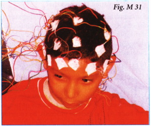
Small, non-invasive electrodes (usually 16 to 32 in number) are placed upon a person's scalp, after careful measurement by a trained technologist, with paste or a glue-like substance to hold them in place. Low voltage signals (5-500 microvolts) are amplified by the EEG machine and stored digitally. The resulting polygraphic display, looking much like a multiple channel seismograph, is typically read by unaided visual inspection. Electrical signals produced by the brain neurons are picked up by the electrodes and transmitted to a polygraph, where they produce separate graphs on moving paper using an ink writing pen or on a computer screen, (fig. M 31)
Procedure
Lie down on the examining table while electrodes are attached to the scalp. Relax whole body and lie first with your eyes open, then later with closed eyes. One may be asked to breathe deeply and rapidly or to stare at a flashing light - both of these activities produce changes in the brain-wave patterns.
The names of the electrode sites use alphabetical abbreviations that identify the lobe or area of the brain to which each electrode refers:
| F | = | frontal |
| Fp | = | frontopolar |
| T | = | temporal |
| C | = | central |
| P | = | parietal |
| O | = | occipital |
| A | = | auricular (ear electrode). |
The localization of the brain waves within the brain regions or lobes is further narrowed by adding electrodes, which are given numbers such as T3, T4, P3, P4. Even numbers identify electrode positions on the right side of the head, and odd numbers refer to the left side. The label V points to electrode sites in the midline of the head. For example, Cz refers to the midline central region of the head.
During an EEG, typically about 100 pages of activity are evaluated. Special attention is paid to the basic waveforms, but brief bursts of energy and responses to stimuli, such as light, are also examined