 Jethalal S. Zaveri
The Skin
The 206 bones of the average adult skeleton work together with the muscles and connective tissues to move, support and protest the vital organs making up the human body. It enables us to run, jump and control overselves with a freedom unknown amongst our mechanical creations. Each individual bone is carefully designed to fulfil its role. Ranging in size from the powerful thigh bone (femur) about 50T mm long and 25 mm across—to the smallest of the wrist bones found at the base of the little finger and shaped like a split-pea. The former must withstand 1200 lbs per square rich during walking; hence it is filled with crosshatching internally for great resistance to pressure. Protection of the brain is one of the most important functions. The skull is made up of eight pieces of bone.
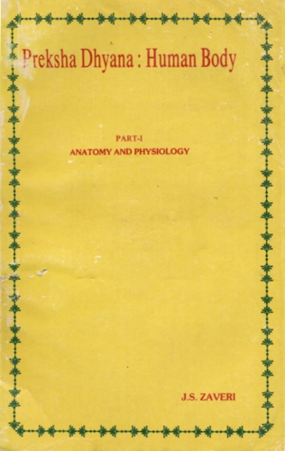
Spine or vertebral column is a marvel of efficient engineering and functionalism. Its vertebrae are formed to compose a cylindrical column strong enough to provide support for the body, yet flexibly articulated to provide for bending, stooping, twisting and a variety of other body motions.
An intervertebral disc of compressible fibrous cartilage is sandwiched between each two adjacent vertebrae cushioning the joint. With this structure, the vertebral column can withstand forces of compression many times the weight of the body.
The rib cage is an excellent example of the versatility of joints and the protective function of skeleton. It protects two delicate organs—lungs and heart, while being able to expand and contract to bring in fresh supplies of air. Cartilage joins the ribs to the breastbone, providing a movable elastic connection. At the back, the ribs are fitted on to the vertebrae by tiny gliding, rotating joints permitting the rib cage to adopt different rhythms of breathing. In contrast to these is the large ball and socket joint of the hip which holds the rounded end of the femur in a self-lubricating socket. The head rotates on the two top vertebrae in the neck enabling us to shake, nod and turn our heads.
Bone Structure
Bone is like reinforced cone rate. Its hardness is imparted by deposits of calcium carbonate and calcium phosphate in the matrix. Strength is imparted by reinforcing fibres. Living cells of the bone are nourished by blood vessels that permeate the bone. Cavities within bones house bone-marrow in which billions of red blood cells and millions of white one are produced everyday.
By three months after conception, a complete skeleton composed of cartilage is formed. Bones are formed later by ossification. Children's bones are more flexible and less brittle and, therefore, less susceptible to fracture. In old age-excessive reabsorption produce osteoporosis and bones become brittle and easily broken.
The Structure of Joints
Our body is in almost constant motion. Legs move in walking and running; hands and arms move in a bewildering variety of activities, jaws open and close; eyes dart back and forth.
We would be as immobile as statues if our skeleton were one continuous structure instead of being composed of more than ¿00 bones connected in joints. The space between the bones in a joint is filled with a lubricating fluid. It reduces the friction between the moving parts. The whole joint is prevented from moving too far by the ligaments which connect the two bones at the joint, so preventing dislocation
Joints are commonly classified as: immovable, slightly movable and freely movable. The last group consists of ball and socket, pivot, and hinge joints. Shoulder joint is one of the most mobile while in the skull flat bony plates are held together rigidly.
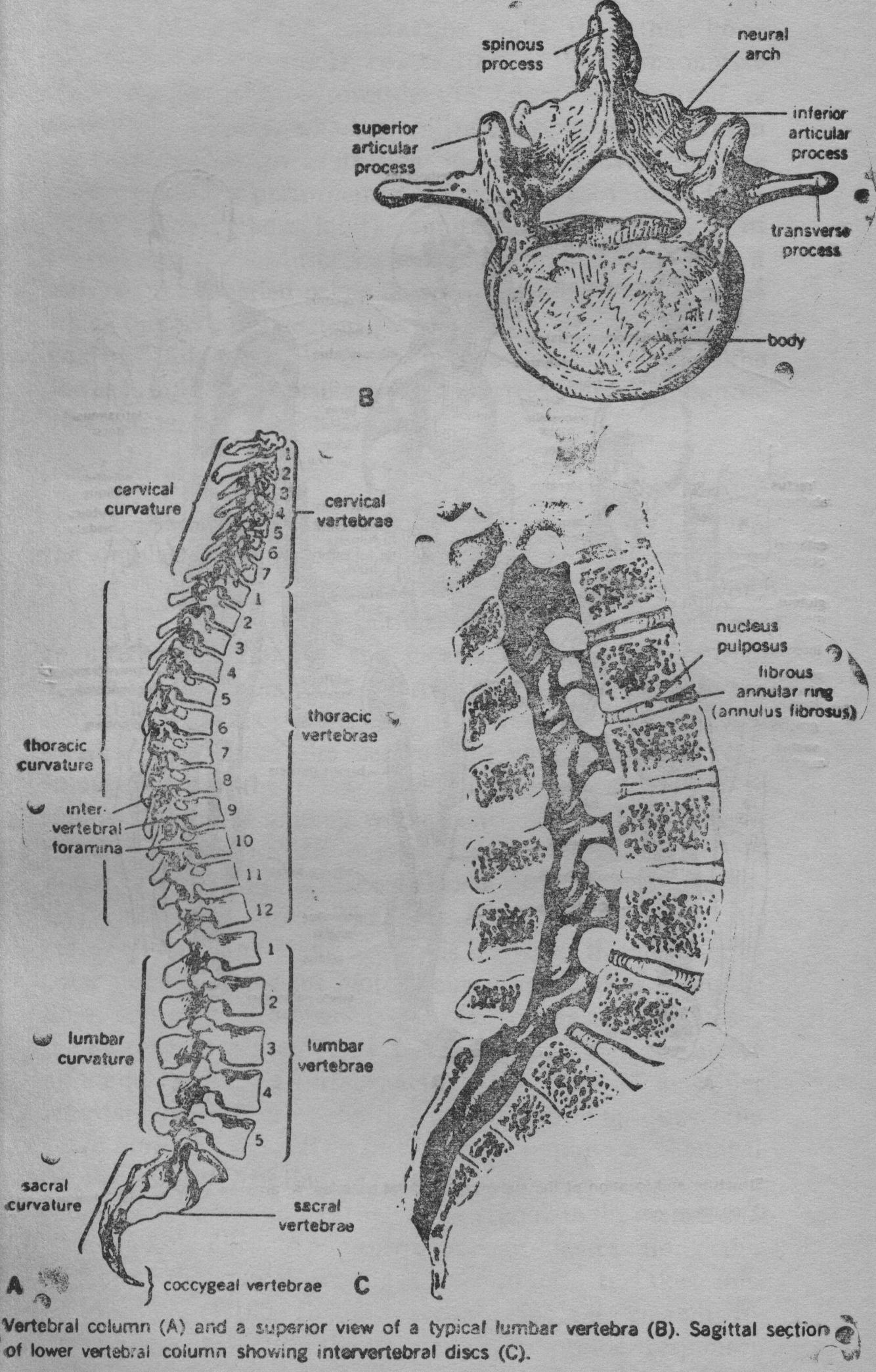
Muscles
Muscles can be called "machines of the body" transforming chemical energy into directed mechanical force and generally producing movement of the body as a whole, of body-parts and of various fluids through specific channels.
In a muscular system makes up 42 percent while in a woman 36 percent of the body weight. It consists of 620 muscles working together to move the 206 bones of the skeleton. A further 30 or so muscles are needed to ensure the passage of food though the intestines, to circulate the blood round the body and operate other internal organs.
There are three types of muscle tissue differing in structure, mode of action and functions:
- Skeletal (or striated) muscles which are used for loco motion. They are attached to bones. They contract rapidly and their contractions permit movement of arms, legs and other parts. They have a capacity for tremendous power but they cannot keep up their exertion long; after a short time they must rest.
- Smooth muscles which line the various organs and intestine. They are slow and steady and can go on contracting day after day without resting.
- Cardiac Muscle (heart) is the special muscle that forms the wall of the heart. They all operate in the same general way because they are specialized in one common property: contractility. They contract and they relax. The fibres which make up the muscles are able to shorten their length by 30 to 40 percent.
With very few exceptions (such as the blinking of the eye) single muscles never contract by themselves; rather whole sets of muscles contract together or in sequence or in opposition to achieve a variety of movement.
Actions of Muscles
Skeletal muscles are firmly attached at each end to different bones. When a muscle contracts, a pull is exerted on both bones but one is stabilized by contractions of other muscles and the contraction pulls the other bone towards it. For example, contraction of the biceps muscle draws the lower arm towards the upper which itself is stabilized. Muscles which stabilize the bone (upper arm in this case) are known as fixation muscles. Movements are complex and the performance of any given movement (e. g. flexing of the elbow joint) requires the coordination of several muscles. Muscles which initiate and maintain a movement are called prime movers or agonists while those which oppose a movement or reverse it are called antagonists. Thus, when the biceps muscle contracts to raise the lower aim, it is the prime mover. The triceps muscle oppose this action and is the antagonist.
The Control of Muscles
To produce the complex movements necessary for even the simplest handling of task, there must be a correspondingly subtle control mechanism. This is the job of the nervous system: it neutralizes the actions of the muscles that are not required and stimulates the muscles which are required. The brain and spinal cord exercise this control through the motor nerve fibres. Each muscle, however, does not have a 'private line' from the central nervous system (CNS). Impulses from the CNS travel down, branching off to supply a group of muscles which contract together, skeletal muscles contract rapidly in response to messages from CNS and develop tension at the same time to produce an effective mechanical force. The area of contact between the motor end plate and muscle fibre is known as the neuro-muscular junction. As the motor neuron approaches a muscle fibre, it branches to form a complex of nerve terminals - the motor end plate. This end plate between the nerve fibre and the muscle surface, acts as a kind of amplifier increasing the effect of the tiny current coming down the nerve fibre. On the arrival of the nerve impulse a chemical (known as acetylcholine) is released from the motor nerve ending and passes across the gap to stimulate the membrane of the muscle fibre. An electric current passes along the surface of the muscle causing it to contract. It takes one millisecond (1/1000th of a second) for the current to pass, the contraction being an all-or-nothing response.
Then the fibre relaxes until another impulse arrives.[1]
Smooth Muscles react much more slowly than skeletal muscles. The nerves when present, alter the activity of the muscle rather than initiating it.
Heart Muscle also has a built-in mechanism to maintain rhythmical contraction quite independently, impulses for contraction coming from within the muscle itself.
The Mechanics of Contraction
Skeletal Muscle fibres contract in response to nervous stimuli and the weakest stimulus that can initiate contraction is known as threshold stimulus. When muscle fibres receive a stimulus strong enough to elicit a response, the fibres contract with the maximum force possible or they no not contract at all. But a skeletal muscle as a whole is made up of millions of individual fibres and, therefore, exhibits graded response rather than all-or-nothing one. Thus the same hand that can pick up an egg without cracking it can also wield a sledge hammer.
The Chemistry of Contraction
The muscles convert chemical energy into mechanical work. Muscles need a good blood supply to bring glucose and oxygen and to remove waste products. The actual chemical process involves the oxidization of glucose, thereby releasing energy which is used by the muscles to contract. The process needs a liberal supply of oxygen. In the absence of oxygen, the muscles can convert the glucose to lactic and which still gives the necessary energy.
Skin (Integument)
The skin is rightly described as the largest functional organ of the human body, since it covers an area of 1.5 to 2 square meters in an average adult person. It is the outer covering of the body and has a wide variety of functions in protecting the watery internal environment of the human body from the ravaging effects of the dry external environment. The skin protects the body from microbes, injury to delicate inner tissues, the injurious effects of chemicals and damage by ultraviolet radiation of the sun. It, thus, is both a mechanical and a chemical barrier between the body and the environment.
Structure of the Skin
Skin is a two-layered covering. The outer layer, the epidermis, is made from epithelial tissue. The thicker inner layer, the dermis is composed of connective tissue. The skin contains, sweat sebaceous glands and hair. It has been calculated that a single square inch (six square cms) of human skin contains an average of 20 blood vessels, 65 hairs, 100 sebaceous glands and 650 sweat glands, 28 nerves, 13 sense-receptors for cold, 78 for heat, 165 pressure and 1300 for pain.
The Epidermis
The outer layer of the skin is the portion of the body one presents to the view of the world. The innermost part of this layer is continually dividing and pushing towards the surface, during which time the nuclei degenerate and the cells die. The skin cells that are visible, therefore, are dead. This outer layer varies in thickness in different parts of the body. It is thickest on the palms and soles. There are neither blood vessels nor nerve endings in this layer.
Some of the cells are specialized for the production of the dark pigment melamine, which is the major determinant of the skin colour. More melamine is produced among certain races than in others and this is the basis of colour difference among races.
Dermis
Dermis, the inner layer, is a thick layer of dense connective tissue. The structure of the dermis contributes great strength, toughness, distensibility and elasticity to the skin.
The dermis contains blood vessels, lymphotics, nerves and some structures derived from the epidermis (known as accessory organs). These are the sweat glands, hair, sebaceous glands and nails.
The upper layer of the dermis contains small projections. Since the epidermis is built on top of these projections, the order layer is also structured in a series of hollows and ridges which change the outer appearance of the skin.
The patterns (called fingerprints) are so characteristic for each individual that they can be used as means of identification.
Accessory Organs of the Skin
Varied functions of the skin are abetted by accessory organs such as hair and nails. More important but less visible are sebaceous and sweat glands.
Hair
No human is totally hairless. A prominent growth is the hair of the scalp. It protects the head and brain proteins from the harmful effects of the Sun's rays and excessive heat. Facial and chest hairs (on males) are signs of sexual maturation. A hair can regrow even if cut, shaved or plucked out. The colour of the hair depends on the amount of melamine present. White hair is the result of the replacement of melamine by tiny air bubbles. Each hair consists of a root and a shaft.
Nails
The nails form a protective covering on the dorsal surfaces of the fingers and toes. Their appearance may give an indication of general body health. Each nail consists of a root and a body. It takes about six months for the nail to grow out from its base to the finger tip.
Sebaceous glands
These are microscopic structures secreting an oily substance—sebum, which forms a protective film over hair and skin.
Sweat glands
These are distributed over most of the body surface but are more abundant in the armpits, the palms, the soles and the forehead.
SWEAT is a clear watery fluid containing 0.5 percent of solids consisting of sodium chloride, other mineral salts and urea. Sweating is one of the mechanism by which the body regulates its temperature. The secretion of sweat is controlled by hypothalamus via the |sympathetic nervous system.
Even at normal body temperature water is lost continuously through the skin as insensible perspiration which evaporates as quickly as it is formed. At environment temperature, above 90° F, the sensible perspiration appears as visible drops of moisture on the skin.
Temperature Regulation
Control of body temperature is very finely balanced in all warm-blooded mammals and it is maintained at an average of 97° C (98.6° F). The body temperature is continuously monitored by the hypothalamus. Thermal receptors in the skin relay information about changes in the temperature of environment to the brain. Various mechanisms keep the body temperature relatively constant even though the skin itself may be quite cool. Skin plays a key role in body-temperature regulation. Heat is lost from the skin surface by radiation, convection and evaporation of sweat. The continuous secretion of perspiration carries off substantial amounts of heat.