Respiration
The body needs a continual supply of oxygen. One may survive for a long time without food, for less than a week without water, but one would not last more than a few minutes without oxygen. In addition, for a continual supply of oxygen, the body also needs some means of disposing of the waste carbon dioxide produced by the function of the body cells. The body also needs an efficient distribution system to deliver the oxygen to all 600 billion body cells and carry off their carbon dioxide. The word respiration is used to describe all the processes associated with the release of energy in the body. Breathing makes a continual replenishment of the oxygen in the lungs, drawing in fresh air from the atmosphere and expels the unwanted carbon-dioxide outside. Blood circulation delivers the oxygen and brings waste gases to the lungs. We have already discussed the circulatory system in the previous section. Here we shall discuss the anatomy and the functions of the respiratory system.
Organs of the System
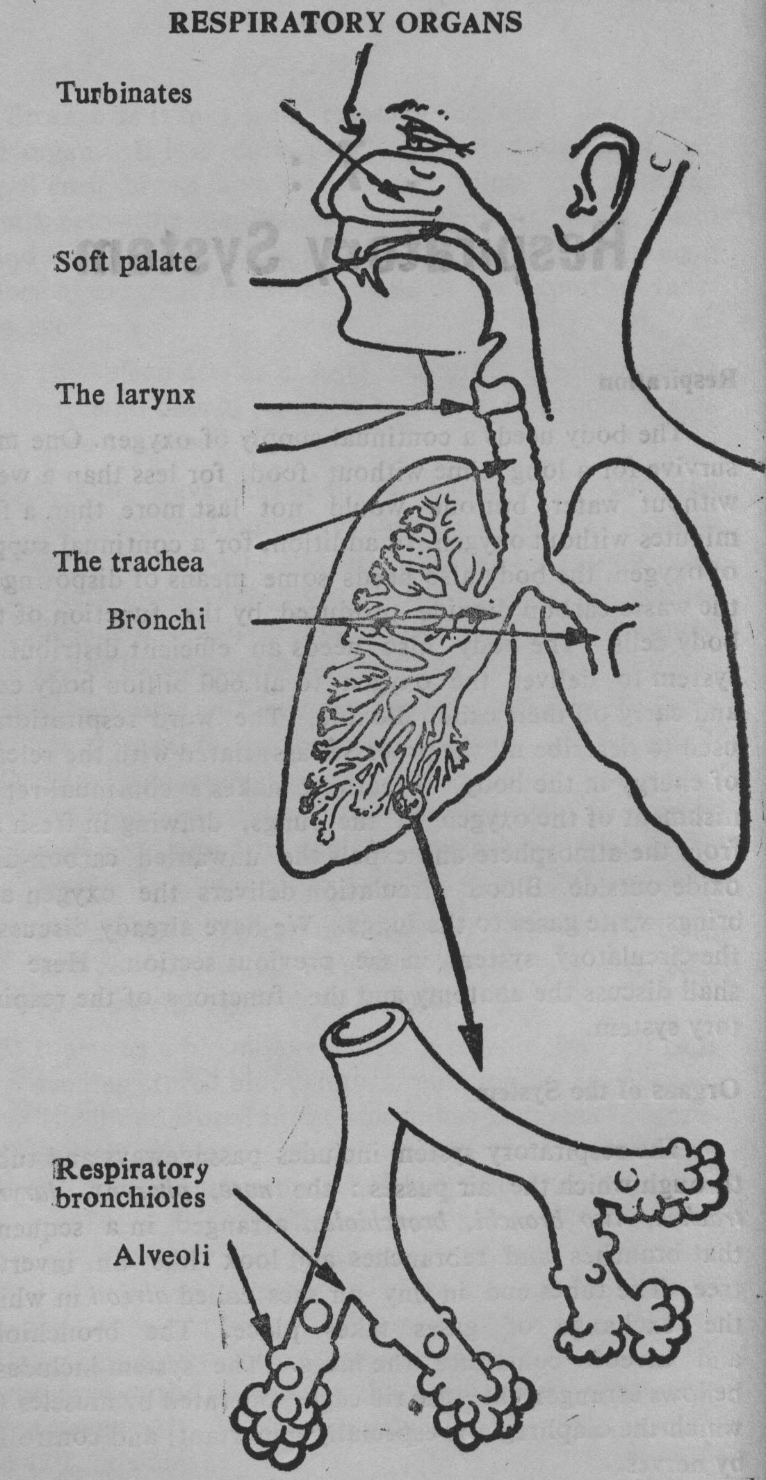
The respiratory system includes passageways and tubes through which the air passes: the nose, pharynx, larynx, trachea, two bronchi, bronchioles, arranged in a sequence that branches and rebranches and look like an inverted tree. The tubes end in tiny air sacs called alveoli in which the exchange of gases takes place. The bronchioles and alveoli constitute the lungs. The system includes a bellows arrangement—the rib cage—operated by muscles (of which the diaphragm is especially important) and controlled by nerves.
The Nose
The nose is the gateway to the respiratory tract. It filters, warms and moistens the incoming air. Nostrils and the area just inside are lined with a picket-fence of short stiff hairs. This is the first line of defence, screening large particles out of the entering air. The interior of the nasal cavity is lined with mucous membrane. Dust and other fine particles and bacteria are caught in the sticky mucus. The mucous membrane also helps to moisten the incoming air. It also contains dense vascular plexus (with copious blood supply) which acts as a radiator to warm the air. The jutting shelves—turbinates—increase the radiating surface making the warming action more effective.
It is also possible to breathe through the mouth but the mouth is not supplied with the apparatus for cleaning, warming and moistening the air. Mouth-breathing, therefore, must be avoided.
The Pharynx and the Larynx
The pharynx is an important junction for the passageways leading into the body. Both air and food have to pass through it. The passage connecting the pharynx with the trachea is called larynx or 'voice-box'. Its main function is the production of speech. But it is also important in breathing and protecting the trachea from foreign objects The epiglottis is a hinged 'trap door' at the entrance of the larynx. During swallowing it closes like a lid to prevent the entry of food into the trachea
The Trachea, the Bronchi and the Bronchioles
The trachea or windpipe is a cylindrical tube about 11 cms. long and about 2 to 2.5 cms in diameter. It lies in front of the aesophagus- Its tubular walls are reinforced by C-shaped rings of cartilage. It divides into two branches - bronchi, one leading into each lung. It is also lined with mucus-secreting cells. Inhaled particles are trapped in the mucus. The cilia (fine hairs) in the lining of the trachea and bronchi whisk up the dust laden mucus upwards towards the pharynx where they can be expectorated. The trachea with its two branches—the bronchi and their numerous branches the bronchial tubes and bronchioles, looks very much like an inverted tree. Each tiny bronchiole terminates in a cluster of minute air sacs, the alveoli which look like a miniature bunch of grapes. An extensive network of capillaries surrounds each alveolus. Gases diffuse back and forth between the alveoli and capillaries network, and the actual exchange of oxygen and carbon dioxide occurs here.
The Chest
The lungs, the heart and major blood vessels are protected by the bony rib cage called chest or thoracic cavity. It is closed at the top by the muscles of the neck; enclosed on the sides, back and front, by the ribs, the vertebrae, and the sternum; is bounded below by the diaphragm, a large sheet of muscle that assumes a dome shape at rest. It is constructed so as to expand and contract readily as the lungs inflate and deflate. The major portion of the chest is taken up by the two lungs. In the centre between the two lungs is situated the heart and major blood vessels.
The Lungs and the Pleura
A pair of human lungs contains about 300 million[1] alveoli covering a total surface area more than 90 sq. mtrs., enough to carpet a tennis court. This enormous surface area provides for an efficient exchange of gases. The two lungs are cone-shaped with the base resting on the diaphragm and the apex into the root of the neck. They are freely movable except at the roots. The right lung is larger and broader than the left, but is slightly shorter. The right lung is divided into 3 lobes and the left one is divided into 2. They are light, porous and spongy. Their internal structure is a mass of branching tubes and air sacs. The alveolus is a roughly globular structure about 100 microns in diameter. In has an extremely thin wall and is surrounded by a network of equally thin-walled capillaries. The total surface area of the capillary network is about the same as that of the alveoli. Each lung is enclosed and protected by a double-walled membrane, the pleura, which also lines the inner surface of the chest wall. A small amount of fluid between two layers acts as a lubricant preventing the lung and chest surface from sticking together.
BREATHING
To a common man respiration is the physical act of breathing in which air is alternately drawn into the lungs and expelled from them. Mostly this is an unconscious act that goes on throughout the day and even when one is asleep In a lifetime, one will take in about 13 million cu. ft. of air. The technical term for breathing is ventilation and it includes two phases: inspiration or breathing-in and expiration or breathing-out. To a physiologist it is divided into: (i) external respiration i.e. passage of oxygen from the lungs into the blood and the passage of carbon dioxide from the blood to the alveoli and (ii) internal respiration in which the body cells exchange carbon dioxide for fresh oxygen carried by the blood. The final aspect of respiration is cellular respiration ie. chemical reactions of oxidation within the cells and release of energy.
Mechanics of Breathing
The lungs may be regarded as elastic sacs the interior of which is opened to the outside via the bronchi, trachea and nose. When the pressure inside is greater than the pressure of the atmosphere, air is expelled from the lungs which is called expiration. When the outside pressure is greater than that of inside, the air flows in. This is inspiration. The pressure differentials are created by the action of diaphragm and other muscles.
The muscles concerned with breathing are (i) the intercostal muscles between the ribs, (ii) the diaphragm between the thorax and the abdomen and (iii) clavicular muscles between the collar bones.
The important mechanism for increasing the volume of the chest cavity is the contraction of the diaphragm. This sheet-like muscle separates the chest from the abdomen. It forms the floor of the chest cavity. It has a dome shape when it is relaxed. When it contract?, it flattens out and descends expanding the chest. In quiet respiration, it moves down about 1.5 cm. producing an increase of about 400 cms3. In forceful breathing the diaphragm may descend as much as 7 cms. producing a far larger increase in capacity.
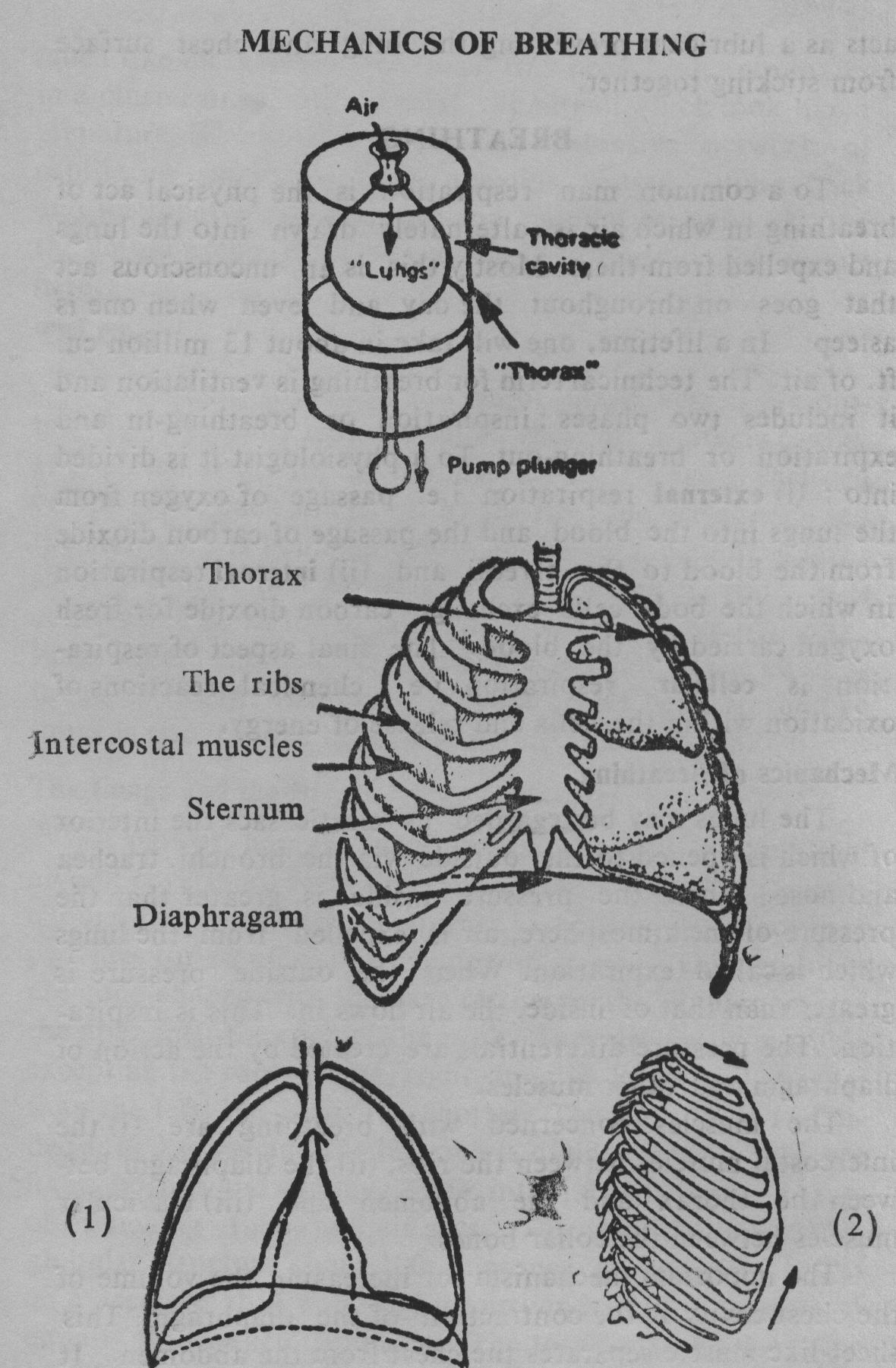
| 1. The depth of the thorax increases when the diaphragm contracts and descends | 2. The intercostal muscles raise the ribs and increase the fore-and-aft diameter of the thorax, and cause the width of rib cage to increase. |
The circumsterence of the chest cavity is increased by another mechanism, the contraction of the intercostal muscles which makes the ribs swing upward and outward expanding the chest cavity. The expansion of the chest cavity automatically inflates the lungs. The third mechanism for breathing is operated by the collar bone. Diaphragmatic breathing is slow and deep; costal breathing is rapid and shallow. At the end of the inspiration the inspiratory muscles relax, the chest volume is decreased and elastic recoil of the lungs expels the air through the passages into the atmosphere.
The total lung capacity is the maximum amount of air the lungs can hold after a maximum inspiration. It is an average of about 6 litres. In a forceful expiration, one can expel about 5 litres in one blow. Even the most forceful expiration does not expel all the air from the lungs. In normal quiet breathing, the volume of air that flows into and out of the lungs with each breath is about ½ litre. Thus, there is normally some air in the lungs after expiration and room for additional air upto 3 litres—after normal inspiration. The volumes and capacities can be modified by breathing exercises and by practising scientific total breathing.[2]
Gas Exchange in the Lungs—External Respiration
The air we breathe into our lungs contains about 21% oxygen and about 79% nitrogen. Small quantities of watervapour, carbon dioxide and other gases are also present. The air we breathe out contains 15% oxygen and 5% carbon dioxide and 79% nitrogen. An exchange of the two gases occurs in the lungs. Oxygen passes out through the thin walls of the alveoli and in through those of the capillaries that surround them. At the same time, there is a net movement of carbon dioxide in the opposite direction.
The factors which favour an effective gas exchange in the lungs are: exceedingly thin walls of the alveoli and capillaries and extremely large surfaces for exchange. The capillary net-work accommodates a large volume of blood - just under a litre at a time. The capillaries themselves are so narrow that the blood cells have to pass through them in a single file; thus each red cell is exposed to the alveola air. Transporting facility is provided by the haemoglobin in the red blood cells.
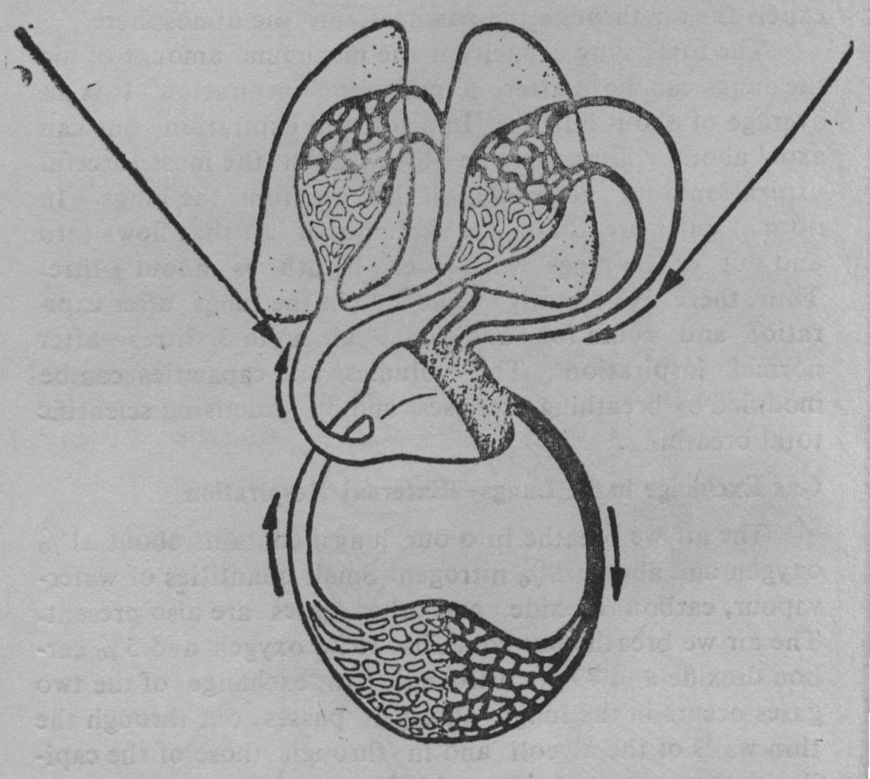
GAS EXCHANGE IN THE LUNGS & TISSUES
| The right side of the heart pump deoxygeoated blood from the body tissues into the pulmonary artery to the lungs. | In the lungs blood is oxygenated. The oxygenated blood flows in the pulmonary vein to the left side of the heart. |
Transport of Gases in the Blood
The blood leaving the lungs is a bright scarlet red. Each haemoglobin molecule can combine (reversibly) with four molecules of oxygen. This oxygenated red blood flows to the heart and from there out into the systemic circulation. There is a continued diffusion of oxygen out of the red cells through the capillary membranes into the issue fluid and from there into the cells.
The transport of carbon dioxide is more complex than that of oxygen. Most of it is carried in the plasma in the form of bicarbonate ions (HC03), some is carried in the form of carbamino haemoglobin and a small amount dissolves in the plasma. Nitrogen which constitutes nearly four-fifths of the volume of air is generally ignored by the body.
Internal Respiration
The major function of the circulatory system is to deliver oxygen and carry off carbon dioxide. The gas exchange between the blood and tissues is very similar to that in the lungs except that the gases go in the opposite direction. Carbon dioxide from the tissues goes into the blood and oxygen from the blood goes into the tissue fluid and from there into the cells.
Control of Respiration
Normally breathing is an unconscious act that goes on continually throughout one's daily activities and even when one is asleep at night. Breathing can also be controlled (consciously) voluntarily to some extent. One can breathe rapidly or slowly, deeply or shallowly at will. One can even stop breathing entirely for a time. But most of the time respiration is under automatic control by special centres in the central nervous system. They are located in the medulla and pons. The medullary respiratory centres set the basic rhythms of respiration. The hypothalamus and cerebral cortex also participate in respiratory control.
The average adult at rest and not emotionally excited breathes about 14 to 20 times a minute. Emotional stimulation, pain, temperature, carbon dioxide level and age cause variations from this basic level. Like the heart-beat, the respiration-rate tends to decrease from the birth to adulthood and increases in old age[3].
In addition to the normal actions of respiration, the respiratory passages participate in a number of reflex mechanisms and other modified movements. Both coughing and sneezing are protective reflex mechanism used to clear the respiratory passages. A cough serves to clear the trachea and bronchi while sneeze serves to clear the passages of the nose and the mouth. A sigh is a deep long-drawn inspiration immediately followed by a shorter but forceful expiration. A yawn is a deep inspiration through a wide open mouth. It produces a more complete ventilation of the lungs than usual.
The estimate of the number of alveoli in an adult human body varies between 250 millions and 600 millions and area 70 to 90 sq. m.
 Jethalal S. Zaveri
