Pranayama is closely associated with our respiratory system. It is the most important system of our body. Our respiration continues involuntarily. Its continuation is life. The moment respiration stops, we die. Pranayama is meant to regulate, control and expand the respiratory system. The theory and practice of Pranayama is incomplete without the adequate knowledge ofthe physiological aspect of respiratory system. Therefore, from scientific point of view, it is essential that we study the structure of respiratory system along with its associated organs.
RESPIRATORY SYSTEM
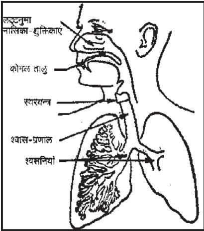
Our respiratory system is mainly a passage for inhalation (intake of breath) and exhalation (outflow of breath). This passage consists of many tubular structures, big and small. It consists of nose—the most exterior organ of the respiratory system—TRACHEA, BRONCHUS and BRONCHIOLE. These are connected with one another in the above sequence. The Bronchiole goes on to be subdivided and becomes gradually smaller in this process. They appear like an inverted tree suspended within the lungs. The BRONCHIOLE end up in the thin-walled air-sacs called ALVEOLI. A group of ALVEOLI looks like a bunch of grapes.
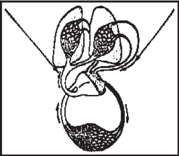
Surrounding ALVEOLI is a complex network of blood capillaries. Air in the form of oxygen and carbon-di-oxide, crossing the walls of capillaries and Alveoli, continues to travel to and fro. So, we see that this is the location where the exchange of oxygen and carbon-di-oxide takes place. Bronchiole and Alveoli together form the inner structure of the lungs. There are no muscles in the lungs. Therefore, the lungs cannot be activated like other muscle driven organ. The diaphragm helps the lungs in the breathing process. Expansion and contraction causes the movement of air in and out of the lungs.
We have already learned that breathing is an involuntary activity which continues to function by itself. But when we practice Pranayama, we are able to regulate the breathing according to our need, to a certain extent. Under these circumstances, it becomes essential that we learn about the respiratory system and its organs in details.
THE NOSE
The nose is the entry point for air. There are fine hair within the nostrils which filter the ingoing air by preventing unwanted materials like dust particles from entering in. The inner wall of the nasal passage is covered with the mucous membrane, a thin film like substance covered with a layer of mucous which does not allow dust particles and other unwanted things to go in. Another function of mucous membrane is to slightly dampen or warm the ingoing air as the need be, before it enters the lungs.
One can breathe by way of mouth also. But there is no filter system here. Neither is there any provision in the mouth to dampen or dry the air before it enters the lungs. That is why it is harmful to breathe through the mouth.
TRACHEA
Trachea is about 11 cm long. Its diameter is 2 to 2.5 cm. It is situated in front of the OESOPHAGUS (the food pipe). When the trachea reaches the lungs, it is subdivided in two parts, one part goes to one lung and the other part to the other. Here this part is known as Bronchus. Inner walls ofbronchi and bronchiole (sub-division ofbronchi) are also covered with mucous membrane. This membrane collects all polluted matter that have entered the lungs along with the inhaled breath. Pores growing inside the trachea and bronchi collect all such dust particles and along with the spittle these are ejected through the oesophagus.
LUNGS
Two lungs are situated within the ribcage. This bone structure protects the heart, Aorta and other arteries. On the upper side of the lungs are the beck muscles and ribs on both sides. At the rear is the spine. In the front is the rib cage. On the lower side, it meets the diaphragm. The diaphragm is a convex, muscular wall. It is situated between the thoracic cavity and the cavity at the top of the stomach. The structure of chest enables it to expand and contract along with these functions ofthe lungs. The lungs take up the most ofthe chest area. In the central region of lungs is the location of heart and other primary arteries. In both the lungs together, there are 300 to 650 million ALVEOLI. Their aggregated area is about 90 square meters, which is roughly as much as the area of a standard tennis court. Since Alveoli can expand to this extent, such a great amount of air can get diffused and circulated in such a small lineal area.
Lungs are shaped like rubber balloons, i.e. they are conical in shape. Base of lungs is at diaphragm. Upper end touches the lower part of neck. Except the lower end, other parts of lungs are capable of movement. The right lung is slightly bigger and broader than the left lung, but is shorter than the left lung in length. Right lung is sub-divided into 3 parts, whereas the left lung has only 2 sub-divisions.
Lungs are light-weight and spongy. Their construction takes innumerable branches of air sacs (Alveoli) and tubular structure called (Bronchiole). Diameter of each Alveolus is approximately 1000 microns. ALVEOLI are round shaped structures. Their inner wall is extremely thin. CAPILLARIES also have similar kind ofthin inner walls. The surface area of ALVEOLI and CAPILLARIES is roughly equal.
BREATHING
In this activity, air is inhaled in the lungs and then exhaled. This activity continues by itself. It continues even when we are sleeping irrespective of day or night. One person in his whole life-time inhales approximately 130 million cubic ft of air. The air that we inhale consists of 21% of oxygen and 79% of nitrogen. Water vapour and carbon-di-oxide are also present. The exhaled air has 15% oxygen, 5 to 6% carbon-di-oxide and 79% nitrogen. There are two factors which influence the diffusion of air in the lungs:
- Extremely thin inner walls of Alveoli and Capillaries.
- Eexpanded surface area of the location where diffusion of air takes place.
In the capillaries surrounding the Alveoli, supply of approximately 1 litre of blood is maintained all the time. Capillaries are so narrow that red-blood corpuscles have to travel in a single file while traversing them. In this way, every red-blood corpuscle has to pass through the layer of air, thereby establishing a close and direct contact with it. Transportation of oxygen is carried out by haemoglobin which is ever present in the red blood cells. Enriched with oxygen, bright-red blood is transported from lungs to heart and then onwards through the blood circulation system to every part of body.
Veins of body bring oxygen-less blood into the right ventricles of the heart and from there it is pumped into the lungs by pulmonary veins. In lungs, blood receives oxygen. Now blood enriched with oxygen is sent to the left ventricle of the heart.
Transportation of carbon-di-oxide is more complicated than that of oxygen. A large amount of carbon-di-oxide is transported in the form of carbonate in veins. Some portion of carbon-di-oxide gets dissolved there itself. 4/ 5 of the inhaled air is nitrogen and body generally ignores it.
From the physiological point of view, respiration is divided into two sections, external respiration and internal respiration.
In the activity of inhalation and exhalation, three muscle-groups participate. It will be suitable to describe them one by one as follows:
- Diaphragm
- Intervascular Muscles
- Thoracic Muscles.
- The Diaphragm is in the lower region of the thoracic cavity. When it contracts, expansion of chest increases. When it becomes lax, it takes the shape of a dome and slips downwards. At the time of inhalation, diaphragm moves about 1.5 cm downwards. By this down movement of diaphragm, expansion of chest increases by almost 400 cubic cm. Gradually when inhalation is capable of becoming long as deep, the diaphragm can be pushed down to the extent of 7 cm. In that case, expansion of chest will be much more.
- Interscular Muscle Group: These muscles groups are attached to ribs. When these muscles are contracted, the circumference of the thoracic cavity increases proportionately. The entire rib cage tends to expand outwards and upwards. Thus the chest expands, and then lungs get the facility of yet more expansion.
- Collar Muscles : When the entire ribcage moves upwards, the upper part of the chest gets the opportunity of expanding more than before. This way the lungs get totally filled up with life-giving oxygen.
In the beginning of inhalation the air pressure in lungs and the outer atmosphere is equal to that of 760 mm (millimeter) of mercury. When the chest expands, the air pressure within the lungs decreases by 2 to 3 mm. To make up for this deficiency, more air is sucked into the lungs from outer atmosphere. At the end of inhalation, diaphragm etc. get contracted. The chest also contracts. Internal air pressure increases. Lungs also contract. Finally, through the breathing passage, exhaled air is pushed out.
The maximum quantity of air reaching in the lungs is known as the LUNG CAPACITY or PRANKSHAMATA. This capacity is on an average upto 6 litres. But this capacity is not fully utilized. During respiration, only about 1 to 2 litres of air is exchanged. The main reason is that during respiration, generally the intravascular muscles and collar muscles are used, diaphragm does not move as much. By activating the diaphragm and by using it during respiration, the capacity of air can be increased from 4 to 5 litres. Therefore, ifwe activate all the three aforesaid muscles during respiration, we can utilize the lung capacity to the maximum. To fully utilize the lung capacity and to increase it, the practice of Pranayama is essential from the scientific point of view.
EXERCISE
- Describe the organs of Respiratory System.
- What do you know about the respiration activity?
- How does exchange of air take place in the lungs?